The main X-ray test for gastric cancer is a double gastrointestinal contrast, commonly known as a barium meal. Although the term “barium meal” is familiar to many people, do you know how this test is done and what the results mean?
Preparation for the test
The patient needs to fast (no food or water) for 8 hours before the test.
Patients should first take an oral gas-producing agent before the test begins. Patients also take an oral contrast medium, usually barium, during the exam. If there is intestinal obstruction or a suspected gastrointestinal fistula, the doctor usually does not recommend a barium meal and may choose pantopamine as the contrast medium, so inform your doctor if you have a previous allergy to pantopamine.
If a gastroscopy has been performed within 1 week, the physician should be informed and these patients should try not to undergo another barium meal because of the damage to the gastric mucosa that can occur after a gastroscopy.
Radiation is produced during the examination, and if a female patient is pregnant or is preparing for pregnancy, she should inform her doctor so that she can weigh the pros and cons of the examination and choose the appropriate option.
Examination procedure
The purpose of oral gas-producing agents is to produce gas in the stomach to fill the stomach and facilitate observation. Therefore, the patient should try to control not to burp after taking the oral gas-producing agent.
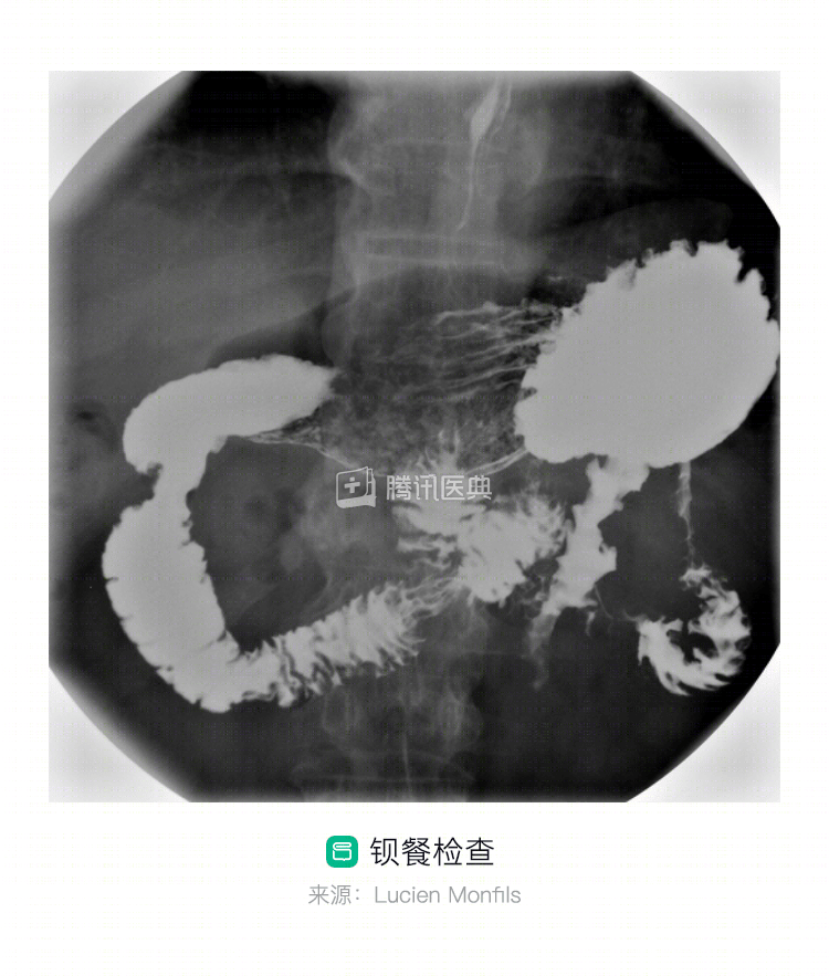
The contrast medium for a barium meal is usually barium sulfate. After the patient enters the examination room, the examiner provides a cup of barium. After standing on the examination equipment, the patient is instructed to drink the barium and rotate his or her body so that the barium is evenly distributed throughout the stomach lining to facilitate the doctor’s diagnosis. The doctor will observe the residue of the contrast agent in the digestive tract and stomach wall through X-ray development to understand the condition of the gastric mucosa. If the examiner finds a suspicious lesion during the examination but it is poorly displayed, the examining equipment may be adjusted to apply local compression to the body surface area corresponding to the suspicious lesion for further observation, such as whether the corresponding stomach wall is stiff and can be displaced, to assist in determining the nature of the lesion.
The radiographs themselves are not unusual for the patient. The contrast solution is usually viscous and less palatable. Some patients may have a bloated feeling after taking the gas-producing agent, which usually resolves on its own.
Post-examination precautions
The barium taken is not absorbed by the body and can be excreted through bowel movements; therefore, patients can drink more water after the examination to facilitate the excretion of the barium.
There will be residual contrast in the gastrointestinal tract after the test, which can interfere with other tests such as abdominal ultrasound, so patients will need to wait for the contrast to pass out of the body before performing other abdominal tests. The doctor will take this into account and arrange the sequence of tests.
How do I understand the test results?
The doctor uses the barium meal to look at the mucosal surface of the GI tract to see if it is intact and smooth, if there is any distortion or disruption of the mucosal fold pattern, and if peristalsis is normal to determine the presence of disease. In the examination report, the following descriptions suggest that the lesion may be malignant:
- Niche shadow irregularity with disruption of the surrounding mucosa. A niche shadow is a manifestation of ulceration or depression of the gastric wall to a certain depth on an x-ray.
- A small nodular filling defect around the niche shadow with a finger pressure sign (irregular curved indentation at the mouth of the niche shadow) or a cleft sign (sharp angle between two finger pressure signs).
- Irregularity at the narrowing of the gastric lumen.
- Stiff morphology of the gastric wall and loss of peristalsis.
- The leathery stomach or leathery stomach. The leathery stomach is the more unusual type of progressive gastric cancer (diffuse infiltrative type), characterized by diffuse thickening and stiffness of the gastric wall, narrowing and poor filling of the gastric lumen, and typical leathery pouch-like changes when extensively involved.
Of course, the presence of the above description is not always malignant, and the doctor will make a judgment in conjunction with the clinical presentation and other test results. (Contributed by Jinyu Huang, Department of Gastrointestinal Oncology, The First Hospital of China Medical University)