Before we answer this question, let’s talk about what exactly PET-CT is.
What is PET-CT?
PET-CT is actually a combination of two tests, PET and a regular CT scan.
CT is an anatomical image that shows the anatomy of the body in cross-section from top to bottom, layer by layer, each layer is only a few millimeters.
PET examination requires injection of a contrast agent into the body, the composition of which is similar to glucose and is labeled with a radioactive substance, so that the metabolism of glucose in the body can be observed. Organs and lesions with high metabolism in the body (including malignant tumors) that take in more glucose will be visualized, which is a functional image.
By fusing the results of both PET and CT, it is possible to see the information of anatomy such as the location, size, shape, and relationship with surrounding tissues of a tumor, and also to distinguish the nature of a tumor by the metabolism of glucose.
For example, if a highly suspicious lung cancer lesion is seen on CT and then on PET it shows a high metabolic sign, like a “burning” flame, then it is likely that the lesion is a lung cancer. Conversely, if the lesion is not found to have much glucose uptake and shows signs of hypometabolism, then the likelihood of malignancy is lower.
Therefore, the significance of the combination of PET and CT is much greater than the significance of both PET and CT alone.
PET-CT diagnoses lung cancer, what does it do?
1.
1. The primary role is to assess the extent of tumor invasion
In other words, it is to rule out the presence or absence of metastasis elsewhere. In the past, when PET-CT was not widely available, the only way to rule out metastases elsewhere was to do head MRI, whole-body bone scan, ultrasound of the abdomen and neck, and so on.
However, even if these tests were done, they only screened the most metastatic areas, not the whole body. PET-CT is the equivalent of the “Monkey King”, which scans you from top to bottom (commonly from the top of the skull to the upper femur).
2. Helps doctors characterize tumors
For lung cancer, the accuracy of PET-CT is about 70% to 80%, which means that 7 or 8 out of 10 people who are diagnosed with “lung cancer” by PET-CT are accurate, which is the duty of PET-CT as a “grassroots court”. This is the duty that PET-CT has to perform as a “grassroots court”. Of course, the final decision has to be made by the “highest people’s court,” which is the pathological examination that has to be done after the lesion is removed.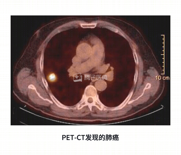
However, pathology testing is necessarily faced with puncture or surgical biopsy. For patients who are older and have a lot of cardiovascular disease, such risks are often unbearable as well. So PET-CT can give a very reliable message about whether to take that risk or not.
PET-CT diagnoses lung cancer, what are the shortcomings?
.
1. The lesions it finds are not always lung cancer
PET-CT has a 20% to 30% error rate, for example, it determines that the tumor has metastasized elsewhere, but then it does surgery and finds that it hasn’t at all.
This is because it is not specific to tumors, but only to organs and lesions that are more metabolically active and can be visualized. Therefore, what appears to be “burning” is not necessarily a tumor. For example, the heart looks like a burning flame flower blooming in the chest, is it a tumor? Of course not. The laryngitis we had in the past, the chronic costochondritis caused by a car accident that bruised our ribs when we were young, the tuberculosis in our lungs, the benign proliferative disease in our abdominal cavity, all of these are not tumors, but they can also show up as “bright” on PET-CT and interfere with our judgment.
2. Not very good at “ground glass shadows”
.
For lung cancer, PET-CT has very low diagnostic validity for small 1 cm lesions, or “ground glass shadows” that are less than 50% solid, and can be easily missed.
This is because these small lesions are the budding stages of lung cancer, which are inherently slow-growing and absorb little glucose, and therefore do not appear hypermetabolic on PET-CT.
3. Cranial imaging to “help”
PET-CT is a poor judge of intracranial lesions.
The limited value of PET-CT in detecting brain metastases from lung cancer is due to the high glucose uptake in normal brain tissue, which causes physiological uptake of 18F-FDG contrast agent to be hypermetabolic and difficult to distinguish from tumors, which requires the use of other PET contrast agents such as 11C-MET.
MRI of the head is commonly used clinically to determine brain metastases.
4. Expensive
Due to medical reform, PET-CT is much less expensive than before, but it is still relatively expensive: it ranges from about 7,000 to 10,000 yuan in various hospitals in Beijing, of which about 2,000 is for medication, which is able to be reimbursed by medical insurance, and the cost of the examination part is not reimbursable. Therefore, doctors advocate selectively recommending patients for this test.
Summary
As a preoperative test, PET-CT has advantages over conventional tests, but it also has “blind spots,” so not all patients will need it, and should be performed according to medical advice. We also need to look at it scientifically and rationally, just because it is “expensive” does not mean that it is “foolproof”. It only provides a reliable basis for puncture, biopsy, surgery, etc. It is our helper, not our only reliance.
Co-reviewed by: Guangdong Provincial People’s Hospital Guangdong Lung Cancer Institute Dr. Chen Zhiyong Dr. Zhang Jiatao
Co-authors: Dr. Wang Xing, Peking University Cancer Hospital