What is thoracic “drainage”? What does it do?
The chest is drained.
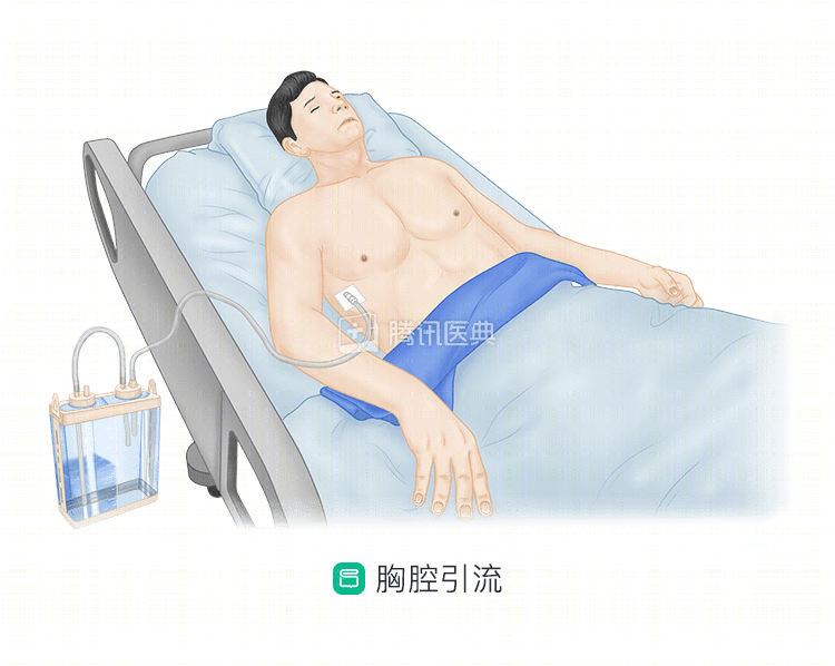
After surgery, the surgeon leaves one or two clear rubber tubes about the thickness of a pencil in the chest wall on the side of the surgery. One end of the tube is inserted into the chest cavity, and the other end is attached to a bottle that holds a certain amount of sterile saline. What is this for?
These are the “drainage tubes” and “drainage bottles” (also called “chest bottles”) that the doctors put in place for the patients. The device is like a window into the chest to drain the fluid and gas. During drainage, blood or bright yellowish fluid flows out of the tube, and a column of water rises and falls in the chest bottle as you breathe. Sometimes when you cough or breathe deeply, bubbles will come out of the bottom of the water column.
In addition, doctors can look through this “window” to see what is going on in the chest cavity, such as whether there is active bleeding, how much bleeding there is, and what needs to be done. The most important thing for the medical staff to see is this window to determine if the drainage is normal and when to remove the tube.
So, what does the drainage “look like”? What are some of the things that might be wrong?
What should I look for in “all kinds” of drainage?
What do I look for in a drainage tube?
The fluid coming out of the chest tube should normally be light blood, becoming lighter and lighter in color over time, and gradually turning into a yellowish, clear fluid.
If the fluid draining out is bright red, thicker, hangs on the inside of the tube, and drains heavily (>50 mL) for a short period of time (half an hour), this is a time to be concerned. This often indicates that bleeding has occurred and the physician will pay close attention and decide whether to go to the operating room to stop the bleeding.
If the color is thick and opaque, as if it has been mixed with milk, this may indicate a “celiac leak,” which is a severe leakage of lymphatic fluid. This may be a sign that you are eating too much, so you can fast and then watch.
If there is any abnormality, such as yellow or green pus or dirty mucus, it may indicate a chest infection, and your doctor will treat it accordingly.
The amount of drainage fluid varies from patient to patient because the procedure varies. In general, the normal amount on the first day after surgery is 300 to 400 mL or less, and too much is indicative of an abnormality. In addition, the drainage volume should be gradually decreasing daily. If it increases instead of decreasing, accompanied by a change in the color and character of the drainage, the physician will further determine and treat it.
To summarize, the color, nature, and volume of fluid draining from the chest tube is a “barometer” that indicates whether the drainage is normal. These conditions are closely monitored by the health care provider. As a patient, your first “job” is to protect the chest tube.
How to protect your chest tube
How do I protect my chest tube?
How do you protect your chest tube?
The first step is to keep the tube and chest bottle closed. Avoid pulling hard on any part of the chest tube or its connections, and be careful to keep the drainage bottle upright and not knock it over.
Second, take care to keep the chest tube open. The tube is relatively soft and easily bent. Therefore, it is important to keep an eye on the chest tube to make sure it is clear when you are wearing a chest strap, dressing, moving around, and sleeping, especially near the body.
Third, keep the chest bottle lower than the chest at all times, whether sitting, lying, or walking, and do not carry the bottle higher to prevent backflow of drainage fluid. You want to get in and out of bed from the side with the chest tube and avoid passing the chest bottle across the bed.
Is the drainage painful? How long does it take to remove the tube?
The doctor will try to use a thinner chest tube to minimize discomfort. However, because the tube is inserted between the ribs and into the chest cavity, and intercostal nerves are abundant, most patients will experience pain, which can be exacerbated by breathing, coughing, or a feeling of “forking”. The pain is unbearable, so you may want to inform your health care provider and use some analgesic medication to relieve it if necessary.
It is normal to be nervous about having a tube in your body and water flowing out of it, and you may be worried about “wanting to get it out sooner or not getting it out. Leave these worries to the medical staff, who are observing the drainage fluid every day and judging the timing of the tube removal with the results of the chest X-ray. In general, the tube should be left in place for 3 to 5 days.
What if the chest fluid doesn’t drain after extubation?
You may also be concerned that:
The chest fluid is not draining.
You may still be worried: Can you remove the tube when 200 mL is still coming out every day? What if the fluid doesn’t come out?
In fact, the human body is an amazing biochemical factory, and there is often a dynamic balance between secretion and absorption of fluid from the chest. The most important thing is that the body has a lot of room to work. But this drainage fluid is actually the good stuff you eat every day, which is rich in high-quality protein, so the loss of drainage fluid is very wasteful. Although foreign scholars generally believe that a chest tube can be removed for a drainage volume of 400 to 500 ml, our research shows that it is safer and more feasible to remove a tube for a drainage volume of less than 250 ml per day. The rest of the fluid will need to be reabsorbed by your body through relentless exercise and increased circulation in an effort to reabsorb it on your own.
In rare cases, the lack of activity and the amount of fluid may cause atelectasis and require a new thoracentesis to release the excess fluid. But remember, the only “secret” that will help you reduce fluid is to be more active. The fastest way to get rid of a chest tube is to be active.
What’s going on with the “fluid” two weeks after extraction?
What’s going on?
Few patients have the problem of “leakage” from the drainage port 2 weeks after extraction when the stitches need to be removed.
First of all, the tightening of one side of the drainage cannula may cause the skin to become purple and even blistered due to ischemia, but this is not a wound ooze, but rather a poorly healed skin.
Also, in general, the drainage tube closes almost completely in 1-2 days without further leakage, bleeding, or oozing, but some patients with diabetes, malnutrition, etc., have slower wound healing, but it usually closes within a week. The actual subcutaneous exudate at the mouth of the drainage tube is sometimes a case of fat liquefaction, which means that the subcutaneous fat is necrotic and the oil flows out of the wound, and this usually happens in obese patients as well.
So when it’s time to remove the stitches, you can consult with your doctor, and generally, the stitches are fine to remove, and if there is an infection or fat liquefaction in the wound, we don’t usually worry about air leaks or seepage inside the chest cavity, we just need to change the wound regularly, and it usually heals quickly again.
Co-reviewed by: Guangdong Provincial People’s Hospital Guangdong Lung Cancer Institute Xie Liang, deputy chief physician Dr. Zheng Shaopeng
Co-authors: Dr. Wang Xing, Peking University Cancer Hospital