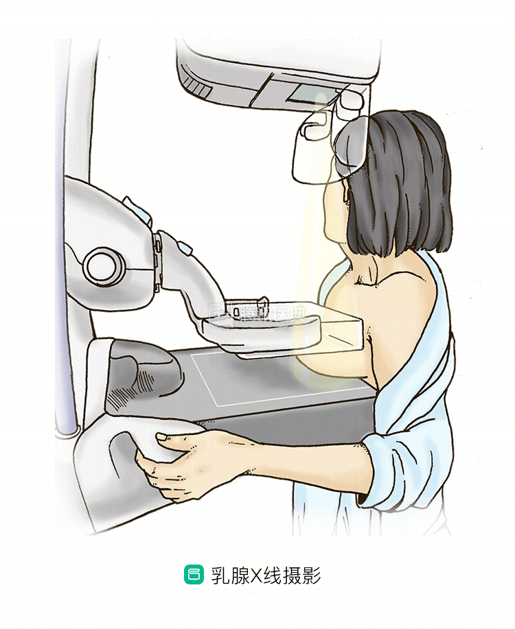
Mammography (MAM), also known as mammography, began in the 1860s with the development of the mammography anode x-ray machine by French physician Gross in 1969.
Since then, the technology has developed rapidly and is now the most important and effective method for diagnosing breast disease, especially for early detection of breast cancer, and is recognized as the first choice for breast cancer screening in many developed countries in Europe and the United States.
What is the use of mammograms?
Mammography is the imaging method of choice for the breast because of its high resolution and good contrast, with a sensitivity of 82% to 89% and a specificity of 87% to 94%.
However, there are limitations to mammography, such as the lack of penetration and density resolution of mammography in dense breasts, which results in insufficient grayscale difference between the lesion and normal tissue and is easily missed. In addition, the need to compress the breast during the examination makes it difficult or impossible to show small breasts and masses close to the chest wall and near the armpit.
Then, along with technological advances, full-field digital mammography (FFDM) was introduced.
FFDM is one of the most widely used mammography techniques in clinical practice, with the advantage of reducing the number of repeat images due to improper technique, improving image contrast, and being sensitive to calcifications. It can detect a proportion of asymptomatic breast cancers, and the examination is inexpensive, simple and convenient.
However, when performing FFDM, the x-ray tube ball remains fixed and only one image is taken per body position, overlapping normal glands and breast lesions can occur, thus impairing the observation of lesion features, especially when the glands are dense, making it more likely to result in missed diagnoses or unnecessary biopsies.
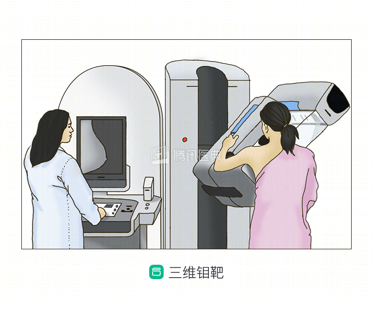
After plain mammography and fully digital mammography, digital breast tomosynthesis, the most advanced digital breast tomosynthesis system (digital breast tomosynthesis, 3D mammography)-was introduced with newer imaging equipment. The use of digital breast tomosynthesis (3D mammography) has significantly improved the clarity of X-ray images of breast cancer lesions.
New screening technology 3D mammography — faster, more accurate, clearer
The digital breast 3D tomosynthesis system, imaginatively referred to as 3D mammography, is an advanced application of flat panel detector-based technology. Rapid image acquisition of the breast through a series of different angles reconstructs an image of the breast at any depth parallel to the plane of the detector, which is further processed to display 3D information.
The first digital tomosynthesis images were first reported in 1997 by Niklason et al. The reconstructed 3D tomosynthesis images of this technique can reduce or eliminate to some extent the influence of normal breast glands on the display of lesions, improve the clarity of breast lesions, increase the contrast between lesions and surrounding glandular tissue, detect lesions more easily, and better show the morphology and edges of lesions, thus improve the detection rate and correct diagnosis rate of breast cancer.
Shortened scan time and effective avoidance of double images
3D mammography requires 5s or less for a total of 10-20 exposures. 3D tomographic images can be acquired not only in the most commonly used internal and external oblique and cephalocaudal positions, but also in other standard projection positions, providing a clearer picture of the breast tissue structure and avoiding overlapping tissue images.
Can be used for dense breasts
Dense breasts have been one of the reasons why non-calcified lesions are easily missed on mammograms. This technique is particularly advantageous in dense mammograms, as it is able to show lesions that may be obscured by normal tissue in conventional radiography.
Increasing breast cancer detection rates
Digital mammography is now widely used for breast cancer screening, but its detection and confirmation rates are unsatisfactory. The overlap of normal breast tissue makes certain signs, such as microscopic masses and calcifications, difficult to visualize, which can affect the final diagnosis.
3D mammography solves some of the key problems of traditional mammography, such as avoiding the discomfort of excessive breast compression and detecting cancerous lesions hidden in overlapping breast tissue. It allows for much better “visibility” of the image, which is advantageous for visualizing lesions, showing lump margins, and determining grading, thereby improving the early detection of breast cancer.
A large retrospective analysis in JAMA found that 3D mammography, when combined with digital mammography, increased invasive breast cancer detection by 41% and increased positive predictive value (PPV) for recall and biopsy by 49% and 21%, respectively.
The technology, approved by the FDA in 2011 and in China in 2014, is available to all women and takes only 4 seconds to examine, especially for women with dense breasts, breast implants, or who have already undergone a biopsy or surgery.
Better accuracy for lesion diagnosis
Researchers comparing 3D mammography with conventional digital radiography have found that 3D has better lesion detection and diagnostic accuracy than conventional digital radiography for masses and structural distortions common to breast disease.
Because 3D viewing removes the glandular component that is obscured above and below the lesion on 2D views, it provides a better view of the lesion and improves the judgment of the nature and volume of the detected lesion.
In addition to the advantages of comparison with conventional 2D images, many authors have compared 3D images with local magnification radiographs and spot compression films added to conventional examinations to show detail, showing that 3D images are superior in the accuracy of lesion diagnosis.
Microcalcifications are often the only manifestation of early breast cancer, and 3D mammography provides tomographic images that exclude overlap of structures such as glands, facilitating the detection of microcalcifications.
What is the future of 3D mammography?
Compared to conventional mammography, 3D mammography provides a clearer view of breast tissue structures while reducing scan time, effectively avoiding overlapping tissue images, and improving the ability to identify benign and malignant conditions in patients with dense glands, detecting small masses that are not detected by 2D mammography due to tissue overlap, and signs that are not recognized due to tissue overlap, in addition to The detection of microcalcifications is greatly improved, increasing the detection of early breast cancer, and is indicated for women with breast implants or who have undergone biopsy or surgery.
As people’s standard of living and health awareness increases, early detection, early diagnosis, and early treatment of breast cancer have become imperative to reduce breast cancer mortality.
3D mammography is highly useful for screening, early diagnosis and preoperative localization of breast cancer. In particular, it is important for the diagnosis of non-palpable early breast cancer.