Radiation therapy (also called radiotherapy), also known as x-ray therapy, involves using high doses of radiation to maximize the killing of prostate cancer cells or to stop their growth and division while minimizing damage to healthy cells.
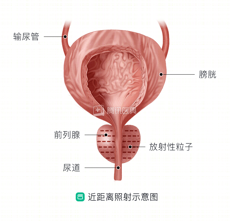
Radiation can be emitted by an external instrument (external irradiation) and used to irradiate the prostate directly; or materials that can produce radiation (radioisotopes) can be implanted through a thin plastic catheter at the site of tumor involvement (internal or brachytherapy), either temporarily (which is removed when the right dose is reached) or permanently.
Radiotherapy procedure
External irradiation needs to be performed at regular intervals over approximately 5 to 8 weeks (usually 5 days per week). For each session, the radiologist will help the patient lie on the treatment table and assume the correct position. Once it is determined that the patient is in the correct position, the physician leaves the treatment room and begins administering radiation therapy.
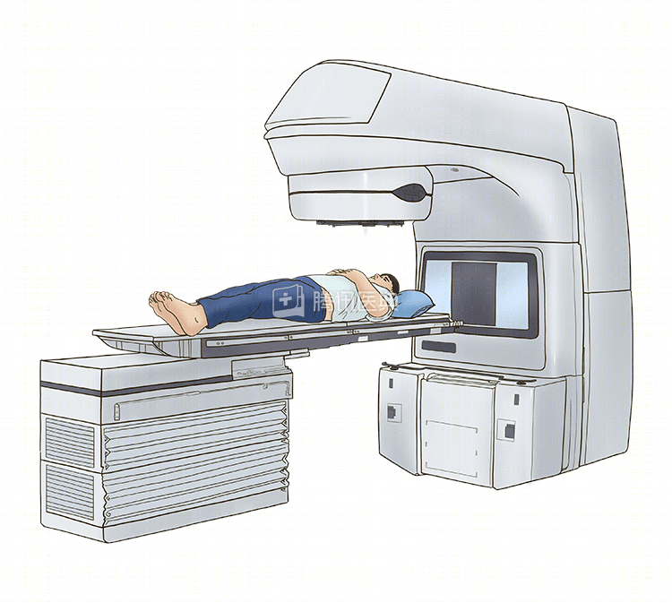
Patients are continuously observed by the physician during treatment. The treatment room is equipped with a video camera and intercom so that the doctor can observe the patient at all times and can hear them. It is important to remain still and relaxed during treatment and to inform the doctor if you have any questions or discomfort.
The doctor will come in and out of the treatment room to reposition the device and change the patient’s position. The device will not touch the patient’s body and the patient will not feel anything during the treatment. Once the treatment is complete, the physician will help them leave the treatment table.
On the first day of treatment and every week thereafter, the radiologist produces a port film to verify that the patient was accurately positioned during treatment.
Shooting field images do not provide diagnostic information, so radiologists cannot learn about disease progression from these images. However, field images are important for radiotherapy, helping to precisely localize the radiation to the tumor site to be treated.
Skin markers
The radiologist will make small freckle-like markings on the patient’s skin along the treatment area. These markings help locate the treatment target and are a semi-permanent outline of the treatment area. Please do not try to wash these marks off; the doctor will re-mark the treatment area if necessary.
Watch your diet
Good nutrition helps counteract the side effects of radiation therapy, promotes recovery, and also fights infection and improves the patient’s quality of life overall. If patients have difficulty eating, they can consult a dietitian to make sure they are getting enough nutrition during radiation therapy.
Related articles: