If your doctor suspects that you have esophageal cancer, he or she may prescribe imaging tests, including chest radiographs and CT. What is the use of these imaging tests? Can they help diagnose esophageal cancer? Here, we will introduce them one by one.
Chest radiographs (i.e., chest x-rays): the old guard is not old, but still an important tool in the diagnosis of esophageal cancer
.
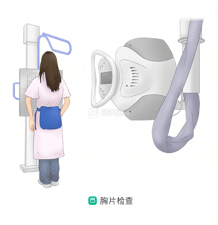
Early esophageal cancer lesions are mostly confined to the mucosal layer, and it is difficult to identify such subtle lesions on X-ray, but experienced doctors will look carefully at the changes in the esophageal mucosal folds and the lumen diastasis, which is still important for confirming early esophageal cancer. The doctor often also uses gastroscopy combined with cytology to supplement the interpretation of X-rays, which can effectively improve the diagnosis of early esophageal cancer.
In clinical practice, doctors often use a “barium meal” (upper gastrointestinal imaging) to enhance the clarity of the esophageal x-ray.
In mid to late stage esophageal cancer, chest X-ray can reveal some abnormalities, such as: thickened post-tracheal soft tissue shadow due to thickened esophageal wall; mass shadow behind trachea and heart due to esophageal mass; dilated middle and upper esophagus due to tumor, etc.
Distant metastasis occurs in esophageal cancer, such as metastasis to the lung, pleura, mediastinal lymph nodes, etc. Chest radiography may reveal pulmonary nodules, pleural effusion, and enlarged mediastinal lymph nodes.
CT examination: irreplaceable in the diagnosis and tumor staging of esophageal cancer
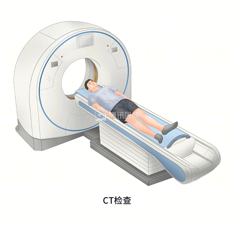
Because the esophagus is surrounded by a layer of fat, CT clearly shows the shape of the esophagus and the relationship of the adjacent organs beneath it. The fat layer between a normal esophagus and adjacent structures is well defined; if CT shows blurred or incomplete boundaries, this indicates the presence of a lesion.
CT examination is a good indicator of the thickness of the esophageal wall, the extent of intramural tumor infiltration, the extent of tumor involvement of surrounding tissues, and metastasis, which is irreplaceable in the subsequent diagnosis and tumor staging of esophageal cancer, especially enhanced-scan CT, which has unparalleled advantages over chest radiography and endoscopy. Sometimes, in cases where barium meal x-ray imaging suspects inoperable resection, a CT scan can be performed to show the relationship between the lesion and the surrounding area to reassess whether surgery is possible.
CT exams are performed on an empty stomach. Before the scan, your doctor may give you an intramuscular antispasmodic to help dilate the normal segment of the esophagus and clarify the extent of the lesion. During the exam, you should lie in a supine position on the examining table, and the doctor will scan your chest and upper abdomen, among other places, continuously. During the scan, you may be asked to swallow 1 to 2 mouthfuls of contrast or air to show the lumen of the esophagus at the site of the lesion.
If needed, your doctor will give you an intravenous contrast and perform an enhanced scan to show the mediastinal vessels and lymph nodes around the esophagus. This is called an enhanced scan CT.
PET-CT: preoperative evaluation to capture surgical indications
PET-CT (positron emission computed tomography) is a combination of PET (positron emission computed tomography) and plain CT scans, with PET providing detailed molecular information on the function and metabolism of the lesion and CT showing the precise anatomical localization of the lesion.
PET-CT has the advantage of not only accurate localization by CT, but also metabolism of each organ or part of the body. By analyzing the imaging information and metabolic data provided by PET-CT, physicians can make more accurate decisions about the diagnosis, treatment, and evaluation of the efficacy of esophageal cancer.
In addition, PET-CT exams can detect distant metastases at other sites.
PET-CT is particularly important in the preoperative evaluation of patients with esophageal cancer, and it is recommended that you refine your PET-CT as much as possible before surgery so that your surgeon can more accurately identify the indications for surgery.