The esophagus is a long, narrow, flexible tube that is an important organ of the digestive system. It is about 25 centimeters in length. The esophagus is responsible for the journey of food from the pharynx into the esophagus, all the way down to the cardia of the stomach, and then into the stomach.
The “one-bite” journey: understanding the function and overall structure of the esophagus
The esophagus is primarily responsible for the “upward and downward” transport of food.
When food is swallowed, the epiglottis (a leaf-like structure at the back of the tongue root) moves back to cover the larynx and prevent food from entering the trachea.
At the same time, the upper esophageal sphincter (esophageal sphincter) relaxes, allowing the food mass to enter the esophagus. The muscles of the esophagus continually contract peristaltically to advance the esophageal mass downward. As the muscles of the esophagus rhythmically peristaltic, the lower esophageal sphincter (low esophageal sphincter) relaxes, allowing the esophageal mass to pass through the cardia and into the stomach.
The “journey” of the esophagus passes through three physiologic strictures, called the “triple strictures of the esophagus” (Figure 1).
- The first stricture is at the junction of the pharynx and esophagus, about 15 cm from the central incisor;
- The second stricture is 7 cm below the entrance to the esophagus, where the left bronchus crosses the esophagus, a place where foreign bodies can easily be retained, about 25 cm from the central incisor;
- The third stricture is at the cleft of the esophagus through the diaphragm, about 40 cm from the central incisor.
These three strictures are clearly seen during a barium meal imaging.
These three strictures, especially the second and third strictures, are frequent sites of esophageal disease (e.g., scarring, contractures, and diverticula) and are often good sites for esophageal cancer. This may be because the physiologic strictures are prone to food retention and become an irritant that predisposes to esophageal disease.
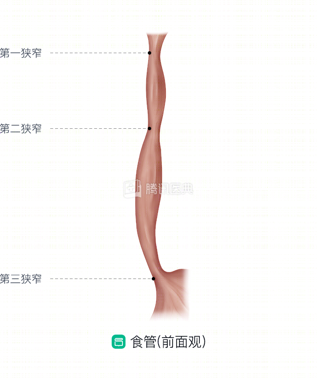
Figure 1
Segmentation of the esophagus
To locate the location of esophageal tumors, physicians often divide the esophagus into three segments: cervical, thoracic, and ventral (Figure 2).
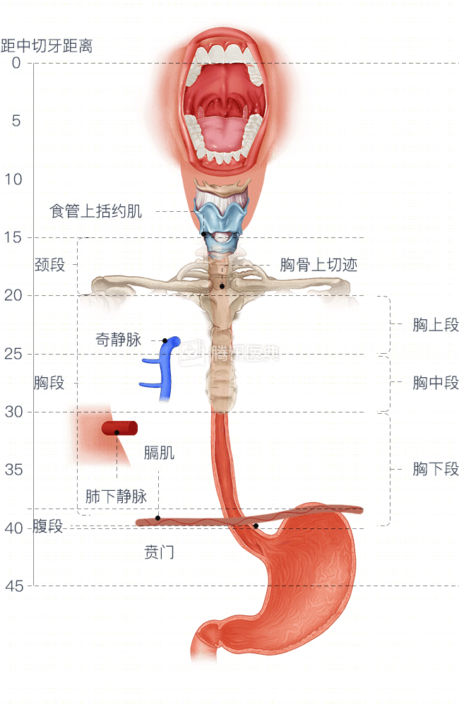
Figure 2
1. Cervical segment of the esophagus
Up to the hypopharynx (upper esophageal sphincter) and up to the entrance of the thorax in the plane of the “suprasternal notch” (Figure 2). The cervical esophagus is like a sentry, once the food reaches the upper esophageal sphincter from the pharynx, it is responsible for receiving the food and informing the following esophagus to go to work.
2. Thoracic segment esophagus
From the upper plane of the sternum all the way to the diaphragm. This segment of the esophagus is the mainstay for transporting food to the next station in a steady stream.
The physician further divided the thoracic segment of the esophagus into 3 segments: upper, middle, and lower (as in Figure 2) for ease of treatment.
- Upper thoracic segment: upper thoracic opening to the level of the lower border of the odd vein arch, about 5 cm long and 20-25 cm from the incisor.
- Mid-thoracic segment: level of the inferior border of the arch of the odd vein to the inferior pulmonary vein, about 5 cm long, 25-30 cm from the incisors.
- Inferior thoracic segment: the level of the inferior pulmonary vein to the cardia, including the esophagogastric junction, about 10 cm long, 30-40 cm from the incisors.
.
3. Ventral segment of the esophagus
A small segment of the esophagus that runs from the diaphragm to the cardia of the stomach. This is the “sprint” force of the esophagus that transports food to the stomach with a final push to open the lower sphincter.
Structure of the esophageal wall
So, what are the upper and lower esophageal sphincters mentioned earlier? This brings us to the structure of the esophageal wall.
The esophageal wall has 4 layers: the mucosa, the submucosa, the lamina propria, and the epithelium (pictured below).
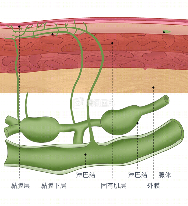
1. The mucosa of the esophagus is covered by squamous epithelium, which is resistant to friction and has a protective effect on the mucosa.
2. The submucosa, which contains mainly connective tissue, as well as lymphocytes, plasma cells, nerve cells, vascular network, and mucus glands, is the “home base” of esophageal immune function, neurotransmission, and blood exchange. The secretion function of the esophageal glands can lubricate the esophageal wall, facilitate the transport of food mass, and resist the small amount of refluxed stomach acid.
3. The muscular layer of the esophagus is made up of two types of muscles, transverse and smooth muscle. The muscle tissue can contract and act as a “conveyor belt” to help push the food mass down. The esophagus is surrounded by two muscle rings at the top and bottom, the upper and lower sphincters mentioned earlier.
4.
4. The epiglottis is the outermost layer of the esophagus, containing larger blood vessels, lymphatics, and nerves, and is also responsible for connecting to the organs surrounding the esophagus. However, the esophagus is only covered by the epithelium in the ventral segment. This makes esophageal cancer more likely to spread; it is also more difficult to repair if perforation or rupture occurs.