There are two main tests that need to be done before surgery:
One is a test to clarify the tumor stage, which is used to measure the severity of the tumor and thus decide whether to operate directly, or to first administer neoadjuvant radiotherapy before surgery, or to administer radical radiotherapy before elective surgery;
The second is a test to assess surgical tolerance, which assesses the function of the heart, lungs, liver, kidneys, and other vital organs to determine the safety of undergoing surgical treatment.
Next, we talk specifically about what is included in these two tests.
Tumor staging examination
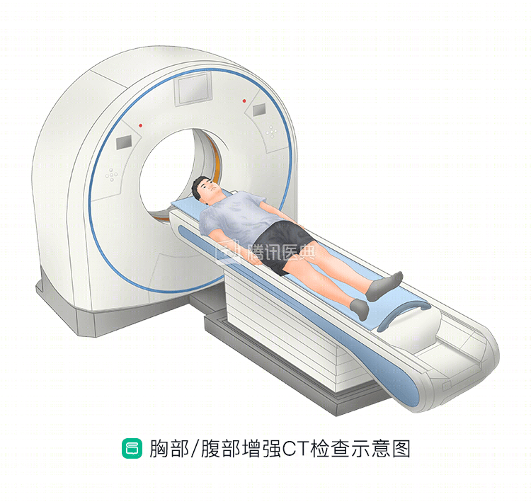
Chest CT
To evaluate the esophagus and stomach, the relationship between the lesion and its surrounding organs and tissues, and to help assess the lymph nodes.
Sometimes, the physician may also prescribe a chest/abdominal enhancement CT, which uses a CT scanner to observe the dynamics of the enhancing contrast in the body and to assess whether the intrathoracic esophageal lesion is invading outward and the extent of involvement of enlarged lymph nodes in the chest and abdomen.
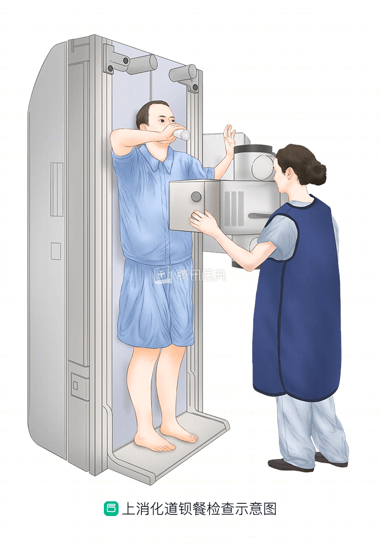
Upper gastrointestinal angiography
You will need to take a “barium meal” where the doctor looks at the abnormal x-ray presentation of the barium as it is imaged along the esophageal wall to assess the esophagus and stomach as a whole (especially longitudinally) and the filling of the stomach to help determine if there is a perforation or fistula.
Whole body PET-CT
Metabolic tracers from positron emission computed tomography (PET) are combined with CT scans to evaluate the esophagus and stomach as a whole, as well as the relationship between the lesion and surrounding organs and tissues, and to assess the extent of esophageal lesions and even distant metastases throughout the body (skull base to hip range human torso) that are suspected. It helps to assess the presence or absence of lymph node metastases and metastases to other organs throughout the body and to refine the clinical staging.
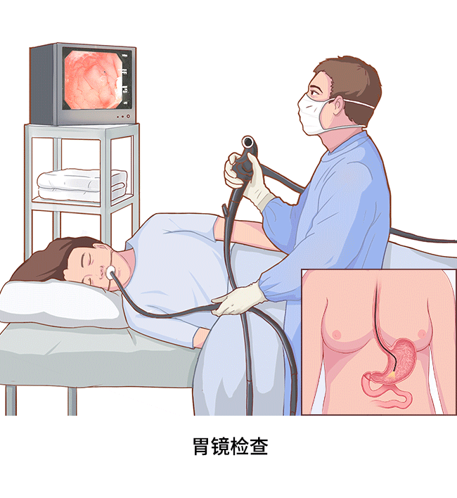
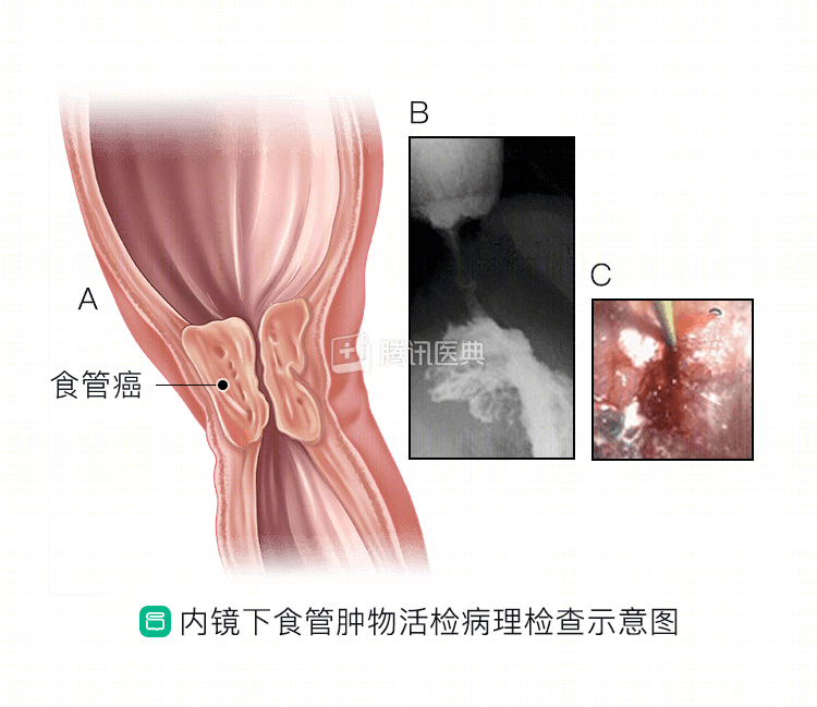
Gastroscopy and pathologic biopsy
The doctor passes a slim tube scope (“endoscope”) with a camera and light source at the end through your mouth and into your esophagus to look at the mucosa in the lumen of the esophagus. If necessary, the endoscopist can also use the endoscope to take a small sample of tissue directly from the esophagus and have a specialist pathologist look at it under a microscope to look for the presence of cancer cells.
It is worth emphasizing that endoscopic pathologic biopsy is the only way to determine if you have esophageal cancer and is one of the main ways to determine the degree of progression of esophageal cancer.
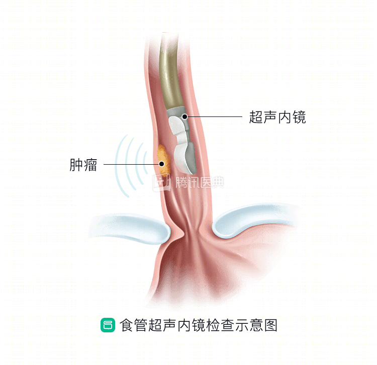
Ultrasound gastroscopy
The physician passes an endoscope with an ultrasound probe on the end, through the mouth and into the lumen of the esophagus, to examine the depth of lesion invasion. It can be used to assess the depth of lesion invasion of the esophageal wall, as well as the condition of the lymph nodes around the esophagus and stomach.
Bronchoscopy
For lesions located above the level of the ramus (primarily the mid-esophagus), assess for invasion of adjacent organs; also indirectly assess for the presence of laryngeal recurrent nerve palsy by looking at the position of the vocal cords.
Cervical and abdominal ultrasound
To assess the lymph nodes in the neck and abdomen and the presence of metastases to other organs.
Surgical tolerance test
Includes tests used to assess the function of individual organs and organs, commonly: electrocardiogram, ambulatory electrocardiogram, cardiac ultrasound, pulmonary function tests, liver and kidney function tests, etc.
If you have underlying conditions such as hypertension, coronary artery disease, diabetes, cerebrovascular disease, or kidney disease, your doctor will need to evaluate your condition before surgery to determine whether you can tolerate the procedure and minimize the risks involved in the procedure.
Here, we would like to highlight the following tests:
24-hour ambulatory electrocardiogram and cardiac ultrasound
It is primarily used to assess the pumping function of the heart to detect potential surgical risks.
Pulmonary function tests and arterial blood gas analysis
Preoperative assessment of your ventilatory function or oxygen diffusion function to determine whether you can tolerate thoracic surgery by means of a blowing lung function assessment and arterial blood gas analysis.
Routine blood tests and biochemical tests
Blood tests for white blood cell, red blood cell, platelet, and hemoglobin levels, liver and kidney function, and potassium, sodium, chloride, and blood glucose.
Systemic nutritional status assessment
Nutritional status is an important indicator to assess a patient’s ability to undergo surgical treatment.
Some data suggest that more than 50% of patients with esophageal cancer are malnourished, and they have a relatively higher mortality rate after surgery. Your doctor will ask you about your diet, weight changes, and general signs over the last 3 months, and will assess your nutrition through blood tests. If there is significant weight loss over a short period of time (more than 10% of your usual weight), your doctor will treat you with preoperative nutritional support.